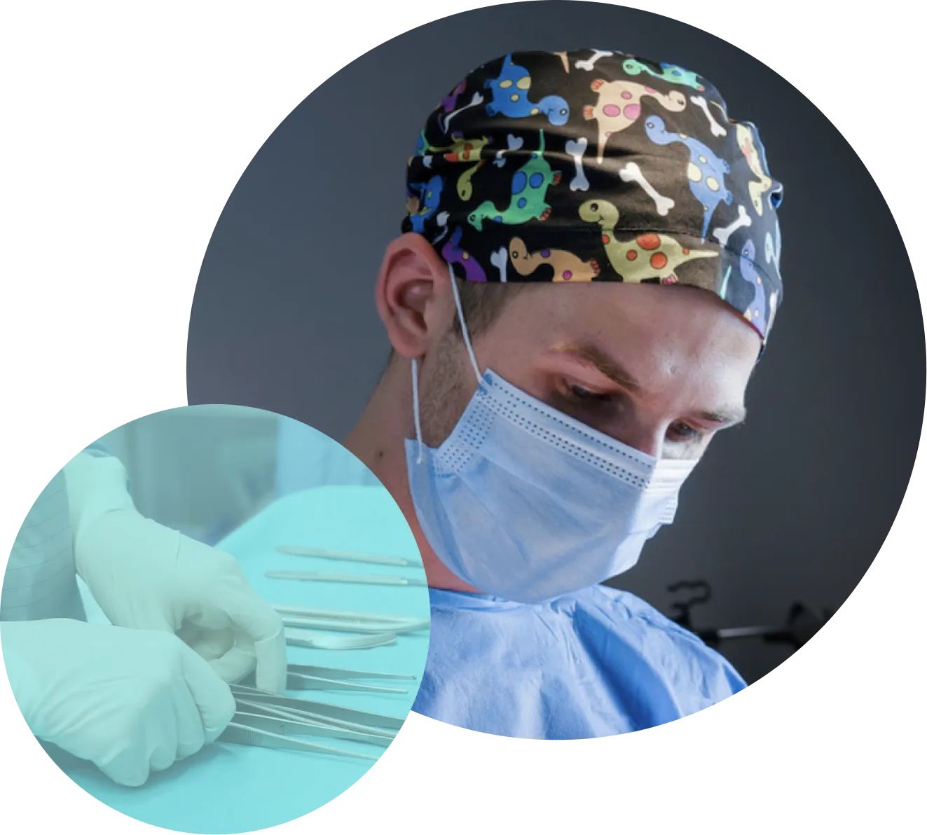
We specialise in diagnosing skin lesions
Since 2019 we have concentrated on a single field. We examine skin lesions, document them, remove them and provide histopathology — and, where needed, an oncology consultation.
From assessing the lesion to excising it and receiving the histopathology result — all in one place.
We specialise in the diagnosis and treatment of skin lesions. Surgical excision preserves tissue for histopathology, which is why we choose it wherever the appearance of a lesion raises doubts.
Others offer surgical mole removal as one of their many services.
We specialise in this.

We look after the whole process — from qualification and the procedure, through histopathology of every surgically removed lesion, to a result accompanied by a clear explanation from a doctor. If the diagnosis calls for further care, we provide a consultation with our oncologist.
We look after the whole process — from qualification and the procedure, through histopathology of every surgically removed lesion, to a result accompanied by a clear explanation from a doctor.
Since 2019 we have concentrated on a single field. We examine skin lesions, document them, remove them and provide histopathology — and, where needed, an oncology consultation.
We created our skin lesion diagnostics programme ourselves and obtained accreditation for it from the Supreme Medical Council of Poland. Only doctors practising at Twoje Znamiona take part in it, which is how we work to one diagnostic standard.
We built our own software, which links your examination result, your medical records and the next stages of care. The system runs only in our centres, so this continuity of care is available to Twoje Znamiona patients alone.
During the examination, the doctor's assessment is supported by a certified AI system. If a lesion raises doubt, the doctor sends its image for a structured second opinion within our nationwide team.
If the doctor recommends an earlier follow-up, a visit within 6 months costs markedly less. Price should not be the reason to put off an examination you have been advised to have.
We are building a nationwide network specialising in skin lesion diagnostics and steadily adding new centres. The examination standard, the doctors' training and the way we work are set across the whole organisation — not one practice at a time.
I was pleasantly surprised by how quickly I could get an appointment. The examination was highly professional and thorough, which matters a great deal because I have many moles. The visit took a little over 30 minutes, but it was well worth the time. I would recommend the centre without hesitation.
The booking staff here are very helpful and committed. The doctor who saw me also turned out to be very pleasant and competent. You can certainly rely on their professionalism. Importantly, the mole examination was carried out very thoroughly. I highly recommend this place to anyone who needs comprehensive skin care.
My visit to the doctor was a very positive experience. She turned out to be very pleasant and competent, which made me feel comfortable during the examination. All my moles were examined thoroughly. I have already booked a follow-up examination; I recommend it to everyone.
We ask every patient for a detailed review after their visit.
You choose the service and the time yourself — no waiting for a phone call.
The Mole Screening Centre is at Kawek 2a/2 in Katowice, in the Brynów district. Public car park by the building.
They work to our single diagnostic standard — the one we set out in our own training programme, accredited by the Supreme Medical Council and available only to doctors practising at Twoje Znamiona.
The idea that “moles are better left alone” is a dangerous myth, and one that can cost you your health. Melanoma caught early can be cured completely. Any atypical mole should be removed surgically without delay.
Laser removal is an option if a dermoscopic examination has been done first and your doctor sees nothing that raises suspicion of cancer. It is worth remembering that a mole removed with a laser leaves nothing behind to send for histopathology.
Surgical excision preserves the whole lesion, so it can be sent for histopathology. A laser vaporises the tissue and leaves no such material. That is why we choose excision wherever the appearance of a lesion raises doubts.
The procedure is carried out by a doctor working at our centre who takes part in our own accredited training programme. This is not a cosmetic treatment and it is not performed by a beautician.
Surgical mole removal is a short procedure, needs no hospital stay and usually takes around 20–30 minutes.
The mole is removed under local anaesthetic, so you stay fully awake and comfortable throughout.
Yes. The first lesion is charged at the standard rate, and every further lesion excised during the same visit at the lower rate for an additional lesion. The doctor decides how many lesions can be removed in one visit, taking their size and location into account.
Wear comfortable clothing that gives easy access to the area being treated. Tell the doctor about any medication you take — especially blood thinners — and about allergies, including to anaesthetics. If anything needs to change, the doctor will say so during qualification.
The price depends on the size of the lesion and where it is — smaller lesions, larger ones and those in sensitive locations are charged differently. All the rates are in the price list higher up this page.
No, it is a separate item in the price list, charged for each lesion sent for testing. If the diagnosis requires immunohistochemical staining, there is no extra charge for it. During qualification the doctor will tell you plainly whether the test is recommended in your case.
Not if the procedure was carried out with us — then the follow-up and stitch removal are included in the price. The separate item in the price list is for patients coming to have stitches removed after a procedure carried out elsewhere.
You can see the available slots straight away in the booking calendar, with no phone call and no waiting to be called back. Online booking works around the clock.
How large a scar you are left with depends on the size of the mole and on how closely you follow the aftercare instructions.
It depends on where the lesion was and how the wound is healing — the doctor gives you the date individually after the procedure and books your follow-up visit.
The result is usually available within 14–21 days; if additional tests are needed, it may take longer. The histopathology report comes with a clear explanation from a doctor, setting out the diagnosis and the next steps. If the result calls for further care, we provide a consultation with our oncologist.
While the wound is healing, avoid soaking it, strenuous exercise and swimming pools, and keep the fresh scar out of the sun. You receive detailed instructions from the doctor after the procedure, matched to where the lesion was.
Five features worth checking yourself. None of them settles anything on its own — whether a lesion needs removing is decided by the doctor, based on the videodermatoscopic image.
Imagine two perpendicular lines through the centre of the mole and compare the opposite halves. Do they match in shape, colour and structure?
Most ordinary moles are symmetrical in structure. Asymmetry can be a feature of dysplastic naevi or of melanoma in its early stages. Any asymmetrical mole should be checked in a dermatoscopic examination.
Before deciding whether a mole should be removed surgically or can safely be treated with a laser, it must be examined with a dermatoscope.
The doctor uses the examination findings to choose the appropriate method. Atypical moles need to be removed surgically, while non-invasive methods are reserved for benign lesions. Laser treatment, cryotherapy and electrocoagulation do not leave a sample for histopathology, which is the only way to make a definitive diagnosis.
Videodermatoscopy — computer-assisted mole examination — is currently the best method of preventive screening for dangerous skin cancers. Examining moles with a videodermatoscope is above all about assessing how the moles under observation change, by comparing images taken over time.
The signs of melanoma can be invisible to the naked eye, which is why viewing moles under magnification matters. A specialised camera placed against the skin sends images to a computer screen and provides up to 140× magnification, revealing structures within the mole. An experienced doctor carries out the examination, supported by a certified AI system that provides an additional check without replacing the doctor's judgement.
A CO₂ laser removes the tissue of the lesion point by point, with no scalpel and no stitches. The procedure usually takes 10–20 minutes and is most often carried out under local anaesthetic.
Only lesions that look benign are qualified for laser removal — among others selected seborrhoeic keratoses, skin tags, viral warts and xanthelasma. The laser vaporises the tissue, so no material is left for histopathology; lesions that raise cancer concerns are removed surgically, with histopathology.
After a melanoma diagnosis, the patient may need further procedures, including a wider excision around the original site and a sentinel lymph node biopsy. An oncology consultation is essential at this stage.
During the consultation, the oncologist explains what should happen after a cancerous lesion has been removed, including the necessary follow-up examinations, possible adjuvant treatment and treatment options for advanced melanoma.
Does your skin need diagnosis or treatment? At Twoje Znamiona you can book a dermatology consultation with a doctor who works not only on assessing moles, but also on treating conditions of the skin, hair and nails.
We help patients with problems such as acne, atopic dermatitis, psoriasis, fungal infections, eczema, inflammatory lesions, itching, seborrhoea, nail problems and recurring rashes. During the visit the doctor will assess your symptoms, carry out the diagnostic work and propose what to do next or how to treat it. If what you want is a preventive assessment of all your moles for melanoma, choose the videodermatoscopic examination instead.